Congratulations!
The PhD Award for excellent PhD thesis 2017 goes to
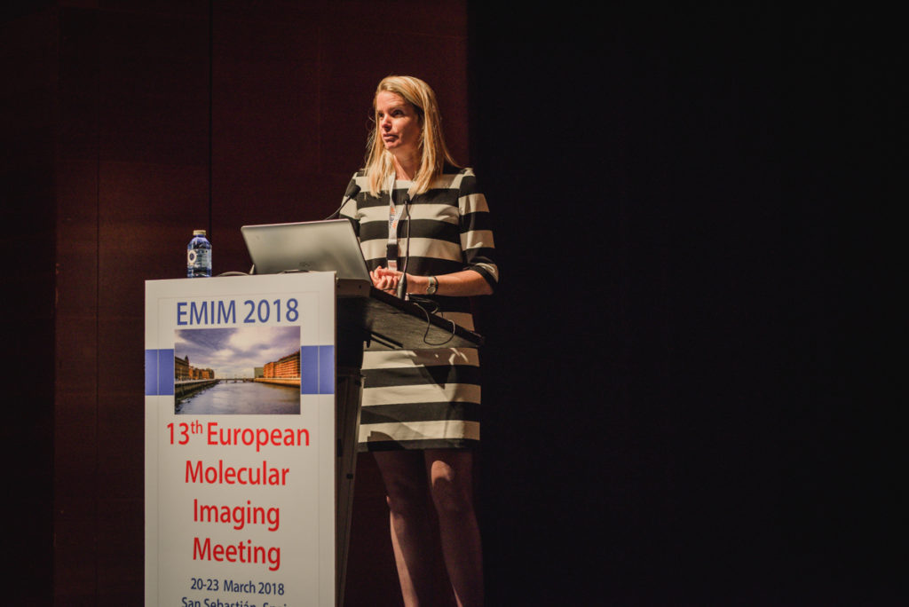
Nynke van den Berg for her thesis on “Advancing surgical guidance: From (hybrid) molecule to man and beyond”
Advancing surgical guidance: From (hybrid) molecule to man and beyond
Abstract
Surgery, often combined with (neo-adjuvant) chemo-, hormonal- or radiotherapy, is considered the main pillar in cancer management. However, during the surgical procedure it is not always clear what exactly has to be removed. My PhD research at the Leiden University Medical Center (dept. Radiology)- Netherlands Cancer Institute/Antoni van Leeuwenhoek Hospital (dept. Urology & dept. Head and Neck Surgery and Oncology) aimed to use interventional molecular imaging technologies to provide the surgeon with additional guidance during surgery as such to improve outcome. We focused on the clinical evaluation and validation of the hybrid, radioactive and fluorescent, tracer indocyanine green (ICG)-technetium 99m-nanocolloid to identify and excise lymph nodes that are in direct contact with the primary tumor (so-called sentinel nodes) in e.g. patients with head-and-neck or urological cancer. Using the hybrid tracer, we created a hybrid approach wherein diagnostic imaging findings could be directly translated into the operation room. Here, for diagnostic imaging the radioactive signature of the tracer could be used to non-invasively identify the target lesions (sentinel nodes). In addition, during surgery, the fluorescent signature of the hybrid tracer allowed visual identification of the sentinel nodes. In comparison to the conventional approach of radiocolloid and blue dye a definitive value of the hybrid tracer was found with more sentinel nodes identified during surgery. This value seemed biggest when sentinel nodes are located near the injection site and/or at locations of complex anatomy (e.g. parotid gland, presacral). Secondly, in collaboration with industrial partners, optimization of radioactive- and fluorescence imaging devices was investigated. Subsequently, in clinical first-in-human pilot studies we found that we were able to improve intraoperative lesion detection and resection. For example, using an optimized fluorescence camera, compared to its predecessor, we could visually more sentinel nodes. Even more so, the use of these optimized fluorescence camera’s resulted in a shift where by the surgeon now can perform real-time fluorescence-guided sentinel node excision rather than using fluorescence imaging for confirmation only.
Impact of work
Radiotracers, in combination with PET or SPECT imaging, have shown to provide a very sensitive technique for the localization of cancer. Furthermore, in combination with anatomical imaging techniques such as CT or MRI, radiotracer imaging has become important for the non-invasive identification of various cancers and detection of metastases, but also for therapy response monitoring.
Surgery, often combined with (neo-adjuvant) chemo-, hormonal- or radiotherapy, is considered the main pillar in cancer management. However, during the surgical procedure it is not always clear what exactly has to be removed. Over the recent years fluorescence imaging by itself has already shown its value for the superficial identification of diseased lesions during surgery. However, fluorescence imaging does not allow for non-invasive imaging and can thus only provide the surgeon with extra guidance when he/she has defined an area of interest.
With the introduction of the hybrid tracer ICG-99mTc-nanocolloid, which is both radioactive and fluorescent we’ve created an hybrid approach in which non-invasive diagnostic imaging can be directly linked to the intraoperative situation. In other words, preoperative non-invasive imaging allows us to identify the regions of interest which can then be evaluated during surgery using the combined use of radiotracing (in-depth localization) and fluorescence imaging (superficial identification). Findings from our sentinel node studies show that using a combination of radioactivity and fluorescence imaging does not only allow the surgeon to plan his/her surgery, but it also has the potential to improve intraoperative lesion identification and resection. This in turn will lead to improved surgical outcome.
We furthermore showed that with the introduction of novel imaging modalities, and/or the improvement of current existing hardware, required for radio- and/or fluorescence-guided surgery a higher detection sensitivity for amongst others the fluorescence signature of the hybrid tracer could be achieved. This means that sensitivity of the camera’s used for surgical guidance play an important role on whether or not a lesion can be detected during surgery. Thus, in order to further refine current surgical procedures we should not only work on the tumor-targeted tracer development site, but also on the clinical hardware.
Nynke’s comments on her future carreer plans
My long term research interest involves setting up a successful translation research line wherein a multidisciplinary approach is taken to bring preclinically developed and validated tracers into the clinic. And to get the beyond “first-in-human” studies. I have a specific interest for radioactive and fluorescence tracers that can help improve 1) diagnosis of cancer; 2) surgical outcome and 3) therapy monitoring. I also realize that in order to perform cutting-edge translational research whereby preclinical developments are translated into the operation theatre, it is essential to build bridges between scientists and MDs. Ideally my own research group would therefor contain various people with different background. Furthermore, I also want to devote part of my time to teaching “future” scientists and MDs as such to decrease the distance between (future) researchers and (future) clinicians, a factor which is now often limiting in (clinical) translational research.

Winner 2017
“The thesis of Nynke reports a ground-breaking approach integrating fluorescence to radioactive signal detection to guide sentinel node surgery in melanoma, oral cavity cancer, penile cancer and prostate cancer. In this respect the thesis discusses the most relevant existing publications on image-assisted surgery in these malignancies and, subsequently proposes a redefinition of the current paradigms in the operating room incorporating a new hybrid approach.
Renato Alfredo Valdés Olmos – Supervisor
The quality of writing of Nynke van den Berg reflects her ability to accurately synthetize both results and discussion for clinicians and researchers in a proper manner.
Nynke is extremely talented and in all these years she motivated clinicians and others researchers in both daily practice as well as in the periodical multidisciplinary meetings in which we had to correlate image findings with surgical outcome.”
“In her PhD thesis, Nynke Van den Berg from the University of Leiden proposed a new integrative molecular imaging methodology combining fluorescence and radioactive detection for image-guided intervention. Her work has been performed at both pre-clinical and clinical levels providing an important breakthrough in the field of cancer surgery. Hybrid fluorescence and radioactive agents have a great potential for image-guided therapy, as demonstrated very nicely by her first-in-man study.
Emmanuelle Canet-Soulas – PhD Award Chair
Besides outstanding publications, Nynke van den Berg pushed the hybrid imaging technology to its clinical application. The PhD award committee is looking forward to follow her scientific achievements in the future and to welcome her at our next ESMI meetings.”
