Intraoperative fluorescence imaging in cancer – Lucia Crane
n the last decades, developments in imaging have led to sensitive imaging modalities that are indispensable in cancer diagnosis and follow-up. A relatively new technique is fluorescence imaging, which has the potential to facilitate intraoperative tumor detection in surgical oncology. Several years of preclinical research have led to the recent introduction of this technique into the clinic. This thesis provides an overview of the technical and biological requirements for intraoperative fluorescence imaging, supported by data from the first clinical feasibility studies in this field, with the emphasis on surgical and gynaecological oncology.
The first part of the thesis focuses on intraoperative near-infrared fluorescence (NIRF) imaging for the detection of the sentinel lymph node (SLN). The standard SLN procedure is based on dual modality detection using radiocolloid and a blue dye, NIRF imaging using indocyanin green (ICG) is a novel method of which the value in gynaecology yet has to be determined. Three clinical studies are discussed, on breast cancer, vulvar cancer and cervical carcinoma, in which NIRF imaging is compared to the standard method.
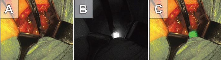
Part II of this thesis focuses on tumor-targeting and tumor-tageted intraoperative fluorescence imaging. As every tumor has a unique biological profile, there is no ‘universal’ target that applies to all cancers. Therefore, target-finding is an issue that needs to be addressed separately for each individual tumor type. Databanks with prospectively collected patient material are essential for biomarker research and translation to the clinic. This is illustrated for ovarian and colorectal cancer (chapters 6-8). The folate receptor alpha is one of the most potent targets in ovarian cancer. This thesis includes a retrospective study on the effect of chemotherapy on FR-alpha expression in ovarian cancer tissue samples (chapter 9). Subsequently, we investigated the value of tumor-targeted intraoperative fluorescence imaging in ovarian cancer patients. To our knowledge, this study is the first to apply tumor-targeted intraoperative imaging in a clinical setting.
Congratulations!
“After my PhD-defence, I have worked in the group of Dr G.M. van Dam for one more year as a postdoc researcher (full-time), focusing on gynecologic and surgical oncology. Currently, I still hold this position, albeit part-time, in which I supervise a number of students who are working on projects in gynecology and neurosurgery. This is combined with a clinical residency in the Department of Gynecology & Obstetrics, in which I work shifts in the outpatient clinic, the delivery room, the general ward and the OR. The combination of clinical work and research allows me to constantly see the bigger picture, clinical relevance and the impact of our scientific work. Furthermore, my critical approach helps me in providing the best patient-tailored care.”
Lucia Crane on her future carreer plans
Together with Angelique Ale Lucia Crane received the first ESMI PhD award for excellent PhD thesis.