PROCESSING BIOMEDICAL IMAGES: Visualisation, Modelling, Segmentation, Quantification, Registration
Images speak for themselves, but exactly what language are they speaking?
On the one hand, incredibly realistic image rendering from the entertainment industry is entering the bio-medical field and will without any doubt change scientists’ and doctors’ practice. On the other hand, progress in image computing science applied to image calculation, analysis and modelling are building up precedently unseen images of biomedical parameters.
TOPIM 2012 brought together prominent specialists from both worlds to discuss the state-of-the-art and the future in modern processing of biomedical images
Topics:
- Image Reconstruction
- Image Processing and Analysis
- Quantification and Modelling
- Image Management and Visualisation
Scientific Committee:
- Bertrand Tavitian – Orsay, France
- Marion de Jong – Rotterdam – The Netherlands
- Adriaan Lammertsma – Amsterdam, The Netherlands
- Boudewijn Lelieveldt – Leiden, The Netherlands
- Renaud Maroy – Orsay, France
- Michal Neeman – Rehovot, Israel
- Vasilis Ntziachristos – Munich, Germany
- Bernd Pichler – Tuebingen, Germany
Programme Specials
The programme started on Monday April 16, 2012 with a three-hour Educational Keynote Lecture given by Adriaan Lammertsma from Amsterdam to enable all participants to comprehend the lectures. This educational introduction was followed by an overview on “Optical Imaging”given by Jorge Ripoll.
We are very pleased and honoured that Terry Jones gave a Key Note Lecture on “The Past Achievements of Brain PET and the Role of Quantification”
“…for my Key Note Evening lecture at the Winter Conference I plan to summarise for each of the application areas what have been the past achievements of brain PET. I hope this will be stimulating for the attendees especially when itemizing the quantitative procedures that accompanied the recording of these achievements. Also, where I can, I will define the experimental paradigms that were used in the studies which should also be of interest.”
Terry Jones
Staffan Strömblad from the Karolinska Institute in Sweden – coordinator of the “Systems Microscopy” FP7 Network – gave a “beyond one’s nose” lecture on
“A systems microscopy platform for studying cell adhesion and migration”
Speakers per (sub-) topic
- Image Reconstruction
Tobias Block – New York, US
Johan Nuyts – Leuven, Belgium
Jorge Ripoll – Heraklion, Greece - Image Processing and Analysis
Daniel Bulte – Oxford, UK
Michal Irani – Rehovot, Israel
Boudewijn Lelieveldt – Leiden, The Netherlands
Renaud Maroy – Paris, France
Jérémie Mattout – Lyon, France
Erik Meijering – Rotterdam, The Netherlands
Sebastien Ourselin – London, UK
Hans Wehrl – Tübingen, Germany - Quantification and Modelling
Heiko Backes – Cologne, Germany
Ronald Boellaard – Amsterdam, The Netherlands
Vincent Cunningham – Aberdeen, UK
Klaus Schäfers – Münster, Germany
Claus Svarer – Copenhagen, Denmark
Paul Tofts – Brighton and Sussex Medical School, UK - Image Management and Visualisation
Wiro Niessen – Rotterdam, The Netherlands
Jason Swedlow – Dundee, UK
TOPIM 2012 was also co-funded by the FP7 projects ENCITE – the European Network for Cell Imaging and Tracking Expertise and INMiND – Imaging of Neuroinflammation in Neurodegenerative Diseases.
TOPIM 2012
THANKS to all of you who contributed to a great TOPIM 2012!
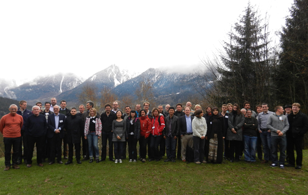
Date: April 15-20, 2012
Place: École des Physique des Houches, Les Houches, France
Meeting documents
Poster Session and Award
Two guided poster sessions chaired by Michal Neeman & Adriaan Lammertsma and by Marion De Jong & Jorge Ripoll were held this year in Les Houches.
The best poster presenters had the opportunity to give a talk the following day. The winners:
Pei Dongfrom Grenoble on
Ralf Engbers from Muenster on

Supported by