THANKS for a great meeting in Dublin, thanks for your talks, posters, your participation and all the discussions – thanks for sharing our vision that interdisciplinary knowledge exchange is the basis for innovation!
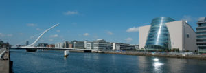
The WMIC 2012 took place in Dublin. The ESMI didn’t organise a separate European Molecular Imaging Meeting that year; the WMIC 2012 was also the annual meeting of the ESMI – the EMIM 2012.
Programme
It is with pleasure that the ESMI treasurer Bernd Pichler from Tuebingen, Germany was the Programme Chair of the congress. He was supported by the entire Committee, and all the chair persons, speakers, and scientists from all over the world who will submit their abstract!
Fabian Kiessling from Aachen, Germany was the Chairperson for the Educational Sessionsin Dublin together with Robert Gillies from Tampa, US. Thanks to all of you for your commitment, efforts, thoughts, and inspiration!
We succeed in attracting high-level experts who are sharing our ambition and helped us to develop an excellent educational concept.
Plenary speaker
- Michal Neeman – Rehovot, Israel on “Multiscale Imaging of the Angiogenic Network.” Michal Neeman is an ESMI Council member read more
- Juhani Knuuti – Turku, Finland on “Imaging of Vulnerable Plaques from
Mice to Man”. Juhani Knuuti is an ESMI Council member read more - Adrian D. Nunn – Princeton, USA on on “Developing high avidity, vascular targeted, ultrasound, molecular imaging agents: Translation to the clinic”
- Ron M.A. Heeren – Amsterdam, The Netherlands on “Image-n-omics: Innovation in Molecular Imaging with Mass Spectrometry”
- Rakesh K. Jain – Boston, USA on “Normalising Tumour Microenvironment
to Treat Cancer: Insights from Intravital Imaging” - Satoshi Minoshima – Washington, USA on “Molecular & Functional Imaging in Neurology – Translational Research and Clinical Applications”
View all abstracts of oral and poster presentations!
Session overview per day
Thursday 6 September 2012
Scientific Session 1: Chemistry & Probes – MRI
Scientific Session 2: Technology & Software – Optical Imaging
Scientific Session 3: Preclinical In Vivo – Metabolic Disease
Scientific Session 4: Translational & Clinical – Oncology
Scientific Session 5: Preclinical In Vivo – Oncology
Scientific Session 6: Chemistry & Probes – Multimodal
Scientific Session 7: Technology & Software – PET/SPECT
Scientific Session 8: Stem Cell, Immune Cell, and Reporter Genes
Scientific Session 9: Preclinical In Vivo – Neurology
Scientific Session 10: Preclinical In Vivo – Oncology
Friday 7 September 2012
Scientific Session 11: Chemistry & Probes – Optical Imaging
Scientific Session 12: Technology & Software – Ultrasound
Scientific Session 13: Preclinical in Vivo – Metabolic Disease
Scientific Session 14: Translational & Clinical – Oncology
Scientific Session 15: Preclinical In Vivo – Oncology
Scientific Session 16: Chemistry & Probes – MRI
Scientific Session 17: Technology & Software – Hybrid Multimodality
Scientific Session 18: Preclinical In Vivo/Translational & Clinical –
Inflammation and Immunology
Scientific Session 19: Translational & Clinical – Neurology
Scientific Session 20: Preclinical In Vivo – Oncology
Saturday 8 September 2012
Scientific Session 21: Chemistry & Probes – Nuclear
Scientific Session 22: Preclinical In Vivo/Translational & Clinical – Cardiology
Scientific Session 23: Preclinical In Vivo – Neurology
Scientific Session 24: Translational & Clinical – Oncology
Scientific Session 25: Preclinical In Vivo – Oncology
Scientific Session 26: Chemistry & Probes – Multimodal
Scientific Session 27: Preclinical In Vivo – Infectious Disease
Scientific Session 28: Preclinical In Vivo – Oncology
Scientific Session 29: CT/US – Technology Methodology – Probes
Scientific Session 30: Preclinical In Vivo – Oncology
Educational Sessions
The WMIC started with the Educational Sessions on Wednesday 5 September 2012.
We have beenm excited to emphasise the importance of the educational part by developing a new concept based on a three year concept developed by outstanding scientists covering the main topics
- Chemistry of contrast media
- What life scientists should know about imaging modalities
- Postprocessing and Cross Validation
- Biology and Pathology
We succeed in attracting high-level experts who are sharing our ambition and helped us to develop an excellent educational concept.
Dublin – the Guiness Storehouse

…thanks for the great party at the “Guinness Storehouse” – Saturday, 8 September
Steering Committee
- Silvio Aime, ESMI President, Italy
- June Key Chung, FASMI President, Korea
- Zaver Bjuwalla, WMIS Vice President, USA
- Juri Gelovani, WMIS President, USA
- Robert Gillies, WMIS Past President, USA
- Clemens W.G.M. Löwik, ESMI Past President, The Netherlands
- Ren-Shyan Liu, FASMI Vice President, Taiwan
- Bernd Pichler, Programme Chair, Germany
Thanks!
View the presentations of the educational sessions
http://www.wmicmeeting.org/wmic-2012/photo-gallery-2012/
Congratulations!
The YIA winner 2012
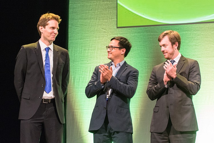
Moritz F. Kirchner – Stanford University on “A Triple-Modality MRI-Photoacoustic-Raman Nanoparticle for pre-and intraoperative Brain Tumor Delineation“
Further Young Investigator Award finalists:
Neal Paragas – on “The Kidney Defends the Urinary System from Infection by Secreting NGAL” – Session 27
Brian M. Zeglis – on “A Pre-Targeted 64Cu-PET Imaging Methodology Based on the huA33 Antibody and Inverse Electron Demand Diels-Alder Click Chemistry” – Session 25
Poster Award winners 2012
per poster walk one poster award has been assigned.
Chairs: Jan Grimm & Claudia Kuntner
Co-Chairs: Frauke Alves, Michael Tweedle, Bruno Weber, Helmut Maecke, Wynne Schiffer, Timothy McCarthy
Preclinical in vivo oncology: P105 In-tumor self-assembly of a caspase-3-sensitive fluorescent probe provides cancer chemotherapy response monitoring. Adam Shuhendler, Stanford University
Preclinical in vivo oncology: P078 Detection of glioblastoma response to temozolomide combined to Avastin® using MRI and PET imaging revealed [18F]-FLT as early predictive markers of treatment efficiency. Aurélien Corroyer-Dulmont, CERVOxy
Neurology: P053 FDG-PET Brain Mapping of Neural Response of American Crows to Human Faces. Robert Miyaoka, University of Washington
Cardiology: P005 Statin therapy improves deep vein thrombosis (DVT) resolution while attenuating thrombus inflammation in vivo: assessment by multiplexed intravital fluorescence imaging. Chase Kessinger, MGH
Infectious Diseases: P037 Whole animal, real-time detection of inflammation in mouse models with conjugated polymer nanoparticles responsive to reactive oxygen and nitrogen species.Adam Shuhendler, Stanford University
Technology & Software – clinical PET, SPECT: P144 A Large Field-of-View PET/CT Scanner for Simultaneous Imaging of Small Animals with Four-Layer Depth-of-Interaction Detectors. Masafumi Furuta, Shimadzu Corp.
Technology & Software – CT, US, Photoacoustics: P176 Quantifying Tumor Interstitial Fluid Pressure in mice xenografts via non-invasive Scanning Acoustic Microscopy. Ralph Pflanzer, Goethe University Frankfurt am Main
Technology & Software -Systems Biology: P167 MRI and NMR study of engineered adipose tissues developed for reconstructive surgery. Marc-Andre Fortin, Universite Laval, CHUQ
Preclinical cell and tissue level – oncology: P323 Three-dimensional visualization of tumor vessel architecture and antibody penetration using multispectral fluorescence ultramicroscopy. Michael Dobosz, Roche Diagnostics GmbH, Technical University of Munich
Preclinical in vivo oncology: P281 In vivo MRI visualization of drug release induced by non focused Ultrasound in an experimental tumor model. Silvia Rizzitelli, University of Torino
Preclinical in vivo oncology: P270 Non-invasive CT-FMT imaging of the biodistribution and tumor accumulation of vascular-targeted polymeric nanomedicines. Sijumon Kunjachan, University Hospital, RWTH – Aachen University
Preclinical cell and tissue level/in vivo – reporter genes, signal transduction & epigenetics: P309 In vivo determination of the fraction of reporter gene expressing cells from a mixed tumor population, using multi-exponential relaxometric MRI. Moriel Vandsburger, Weizmann Institute of Science
Technology & Software – optical imaging: P376 Characterization of Multivalent Targeted Molecular Imaging Probes. Yolaine Jeune-Smith, H. Lee Moffitt Cancer Center & Research Institute
Technology & Software – Hybrid Multimodality: P355 The effects of number of segments in segmentation-based whole-body PET/MR attenuation correction: evaluation with PET/CT data of liver and spine cancer patients. Joong Hyun Kim, Seoul National University
Chemistry and Probes – Nuclear: P227 Synthesis and radiopharmacological evaluation of an 18F-labelled norbornene derivative for rapid copper-free click chemistry reactions. James Knight, University of Alberta
Chemistry and Probes – MRI: P202 Overcoming Biological MT Effects by use of ParaCEST MRI Contrast Agents Possessing Highly Shifted Amide Proton Signals. Mark Milne, Universtiy of Western Ontario
Preclinical in vivo oncology: P545 Detection of Cell Death with GE152 in a Small Animal Model: A Tool for Early Assessment of Tumour Therapy Response. Susan Hoppmann, GE Healthcare
Preclinical in vivo oncology: P554 Different anesthetics impact tumor hypoxia and muscle oxygenation. Moritz Mahling, University of Tuebingen
Neurology: P513 Ultra-Sensitive Molecular Magnetic Resonance Imaging of Cerebrovascular Cell Activation. Axel Montagne, INSERM UMR-S 919
Cardiology: P455 Comparison between magnetic nanoparticle-combined cardiac MRI and conventional cardiac MRI for detection of myocardial inflammation, and visualization of inflammatory evolution in experimental autoimmune myocarditis rat model. Hyeyoung Moon, Korea Science Institute, University of Science and Technology
Metabolic Disease: P592 Manganese-enhanced MRI distinguishes normoglycemic and type 2 diabetic patients. Laurent Vinet, University of Geneva
Translational and clinical oncology: P622 Optical-guided surgery of fibrosarcoma on cat patients. A veterinary clinical study, Christiane Wenk, CRI-INSERM U823, Université Joseph Fourier
Technology and Software – MRI: P583Improved magnetic particle spectrometer providing high field amplitudes for investigation of hysteresis effect in superparamagnetic nanoparticle tracers, Marlitt Erbe, University of Lübeck
Chemistry and Probes – CT, US, multimodal: P418 Synthesis and Application of AOI-Derivatives for in-vivo Imaging of Aβ-Plaques in APP23 Alzheimer Mice with Near Infrared Optical Imaging and MRI, Christian Kesenheimer, Eberhard Karls University Tuebingen