To subscribe to this podcast, copy and paste the RSS URL https://feeds.buzzsprout.com/2045482.rss into the podcast app of your choice.
Edition August 2025
Episode #17 – live from Tuscany:
Rodney Hicks, Melbourne
🎙️Tune in for a dynamic discussion on how NETTER-2 may reshape treatment strategies for neuroendocrine tumours, what it means for the wider field of theranostics, and where radioligand therapy is headed next.
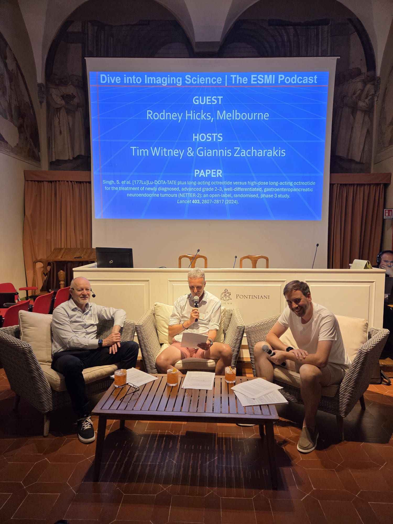
For our seventeenth edition, we are at the stunning Certosa di Pontignano in Tuscany – the home of TOPIM, which focussed this year on Theranostics.
Suitably, we discuss one of the most important recent studies in the field of nuclear medicine and oncology — the NETTER-2 trial, published in The Lancet in 2024. This landmark phase 3 study was the first to investigate radioligand therapy as a first-line treatment in patients with advanced grade 2–3 gastroenteropancreatic neuroendocrine tumours. The results showed a dramatic improvement in progression-free survival and response rates compared with high-dose octreotide, setting the stage for radioligand therapy as a potential new standard of care.
To guide us through this groundbreaking work, we are joined by a very special guest, Professor Rodney Hicks, a global leader in PET imaging and therapeutic nuclear medicine. Over the past three decades, Rod has been at the forefront of bringing molecular imaging and theranostics from the research lab into routine clinical practice. He has played a pivotal role in shaping how PET is used to diagnose, monitor, and now treat cancer, and his vision has helped establish theranostics as one of the most exciting frontiers in oncology today.
Selected publication:
About the Guest
Professor Rodney Hicks is Director of the Centre for Molecular Imaging and Therapeutic Nuclear Medicine at the Peter MacCallum Cancer Centre in Melbourne, Australia.
He has pioneered the use of PET and PET/CT in the assessment of cancer. His group has a strong focus on development of novel theranostic agents and translational research linking imaging phenotype to genotype. He is actively involved in the therapeutic nuclear medicine, especially for the treatment of neuroendocrine tumours and prostate cancer. Rod has published over 500 peer-reviewed articles and more than 20 book chapters and is Editor-in-Chief of Cancer Imaging and International Associate Editor of the Journal of Nuclear Medicine and Lancet Oncology. Read more about his work at Google scholar.
Edition July 2025
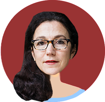
Episode #16: Markita Landry, UC Berkeley
🎙️Tune in for a dynamic discussion on innovative probes work, the function of dopamine in the brain, and how these new imaging tools can be used in the future to reveal new biology.
This episode features a truly special guest: Markita Del Carpio Landry, the inaugural Trailblazer Plenary Lecturer at the upcoming EMIM in Ljubljana next March! !
“The Trailblazer Lecture celebrates a rising leader in imaging science whose trajectory is unmistakably pointing toward lasting impact and excellence at the highest level. Markita is a perfect fit for such an accolade.”
In the Landry lab, they have pioneered synthetic bio-mimetic nanocomposites for fluorescence imaging and targeted delivery of biological cargoes.
We talk with Markita about her career path, about the excitement to develop new imaging tools, and how gaining an academic position gave her the freedom to explore biological questions. We also discuss the importance of collaboration in science.
🎯 The paper Markita selected for this episode, from Yulong Li’s lab and published in Science, features a sophisticated molecular sensor that reads out dopamine signalling in the brain via far-red fluorescence.
The group describes how this innovative new tool can be multiplexed with other fluorescent readouts of acetylcholine and cyclic AMP. Starting in cells, before progressing to tissue sections, zebrafish, and finally in living mice, they demonstrate the ability to detect multiple neurochemicals simultaneously – quite a feat!
 For this episode, we are raising a glass to celebrate the inauguration of the Trailblazer Lectures with a classic: the Champagne Cocktail.
RECIPE: Drop a sugar cube into a flute glass, soak it with a few dashes of Angostura bitters, add a shot of Cognac, then top up with Champagne. It’s a timeless twist on the most beloved celebration drink!
Selected publication:
In vivo multiplex imaging of dynamic neurochemical networks with designed far-red dopamine sensors.
Edition June/2025
Episode #15: Vasilis Ntziachristos, Munich
Tune in for a dynamic discussion on how the interplay of light and sound is revolutionizing our understanding of disease.
🎙️ Welcome to Episode 15 of “Dive into Imaging Science”!
IIn this edition, we’re delighted to introduce: none other than Vasilis Ntziachristos as our guest!
Vasilis is a distinguished leader in the field of optical and optoacoustic imaging, renowned for his pioneering work in fluorescence molecular tomography, fluorescence-guided surgery, and optoacoustic mesoscopy. But his career didn’t begin there. In this episode, we learn how a shift from physics to engineering – and from NMR to optical imaging – was sparked by the influence of key mentors in the field.
With his characteristic charm and charisma, Vasilis reflects on the importance of surrounding oneself with those who challenge boundaries and reimagine what’s possible – an ethos that has defined his own scientific journey.
🎯 Staying close to home, this episode explores how optoacoustic mesoscopic imaging is being used to study colitis, inspired by research from the labs of Sarah Bohndiek in Cambridge and Adrian Regensburger in Erlangen:
Published in Advanced Science, their work introduces a novel ‘transrectal absorber guide’ – an imaging approach that enables visualization of the colon from outside the body. Using this new method, they surprisingly demonstrate how inflammation reduces blood vessel coverage and length but increases vessel diameter.
Vasilis walks us through the fundamentals of this technique, and we dive into the nuances of signal acquisition and image segmentation.
For this episode it was quite easy to choose. Something that takes us back 20 years or so in Boston and the early days of fluorescence molecular tomography, a drink enjoyed at Sonsie, one of the best bars in downtown Boston at Newbury street: The Grey Goose martini – a twist on the vodka martini cocktail
Recipe: 3 parts Grey Goose Vodka, 1 part dry Vermouth and garnish with cocktail olives. Great things can come in very simple forms.

Selected publication:
In Vivo Assessment of Deep Vascular Patterns in Murine Colitis Using Optoacoustic Mesoscopic Imaging
Edition April/2025
Episode #14: Peter J.H. Scott, University of Michigan
🎙️ Welcome to Episode 14 of “Dive into Imaging Science”!
In this edition, we’re thrilled to welcome a very special guest: Peter Scott!
Peter’s passion for radiochemistry and his dedication to translating research into clinical applications have made a lasting impact on the field of Theranostics – also the central theme of this year’s TOPIM (don’t forget to check it out!). By following Peter’s inspiring career journey, we gain valuable insights into how the field has evolved over the years.
The Scott Lab focuses on using PET radiotracers to deepen our understanding of disease mechanisms and to develop companion diagnostics that support therapeutic innovation. A key focus of Peter’s work is the design and synthesis of novel PET tracers for imaging CNS disorders such as Alzheimer’s disease.
🎯 This brings us to this episode’s spotlight topic: Radiotheranostics in Alzheimer’s Disease.
Our discussion is sparked by a recent study from the group of Tara E. Mastren at the University of Utah, published in The Journal of Nuclear Medicine. The paper investigates the use of targeted alpha therapy to reduce Amyloid-β aggregates – an intriguing approach that raises important questions about its potential in Alzheimer’s treatment. We explore the immense potential of radiotheranostics in CNS disorders, as well as key challenges such as delivering compounds across the blood-brain barrier and minimizing effects on healthy tissue.
Continuing our tradition, we celebrate our guest with a special cocktail. This time, a nod to Peter’s love of gin and his British background. For this edition: the “Bee’s Knees”, a classic from the Prohibition era, with a modern twist inspired by The Gin Vault in Birmingham. A simple, elegant sour to keep us energized — and just the right mix of sharp, sweet, and smooth.
Recipe: 3 parts Gin – 1 part lemon juice – 1 part blood orange juice – ½ part honey syrup.
Selected publication:
Development of a 213Bi-Labeled Pyridyl Benzofuran for Targeted α-Therapy of Amyloid-β Aggregates
Holiday/CHRISTMAS Edition 12/2024 
Episode #13: Fabian Kiessling, Aachen
Welcome to this special Holiday / Christmas edition of “Dive into imaging science”! Giannis and Tim have their festive hats on and are wearing their dodgy Christmas jumpers, ready for a cracker (!) of an episode. And we have a very special guest on the show – none other than our current past president, Fabian Kiessling, who heads up the Institute of Experimental Molecular Imaging and the Helmholtz Institute for Biomedical Engineering at Aachen University.
We explore how to ‘make the visible invisible’ in the groundbreaking paper from the lab of Guosong Hong at Stanford by achieving optical transparency in live animals using a food dye that’s an ingredient in many potato chips! We conclude that this isn’t, in fact, the end of non-invasive imaging; instead, it opens a new frontier for optical and photoacoustic techniques.
Along the way, we learn that Fabian could have been a policeman if only he hadn’t been “a little bit weak in differentiating red and green” and that being a scientist is very much like being an artist. We discuss reproducibility in science, the importance of co-locating labs and people, and were swept along by Fabian’s passion for imaging science.
Continuing our tradition, we enjoy a special cocktail :
It’s a Christmas and Aachen special so what better way to celebrate it with a twist on the Gluehwein Cretan style: Giannis’ own home-made brandy and raki (cretan grappa that is), red wine, cinnamon sticks, cloves, honey and a red orange slice from his garden.

Selected publication:
Achieving optical transparency in live animals with absorbing molecules
Ou Z, Duh YS, Rommelfanger NJ, Keck CHC, Jiang S, Brinson K Jr, Zhao S, Schmidt EL, Wu X, Yang F, Cai B, Cui H, Qi W, Wu S, Tantry A, Roth R, Ding J, Chen X, Kaltschmidt JA, Brongersma ML, Hong G. Achieving optical transparency in live animals with absorbing molecules. Science. 2024 Sep 6;385(6713):eadm6869. doi: 10.1126/science.adm6869. Epub 2024 Sep 6. PMID: 39236186.
Episode #12: Elisa Konofagou, New York
Episode #12: Elisa Konofagou, New York
We continue at full speed with back to back podcasts hosting leaders in the field of molecular imaging. In this edition we welcome Elisa Konofagou and share her passion for ultrasound imaging and its unique capabilities for visualising biology inside the human body. Her group has developed methods for estimating minute deformation as a result of physiological function, such as in the heart and vessels, and displacements induced by the ultrasound wave itself, such as in tumours and nerves. She is particularly interested in translating these technologies to a clinical setting and impacting the improvement of healthcare.
The use of innovative ultrasound imaging to solve a clinical problem is the focus of this edition. The discussion revolves around a paper presenting an artful application of ultrasound localization microscopy to image the hemodynamics of myocardial microvasculature in patients. The paper was recently published from the group of Meng-Xing Tang at Imperial College London in Nature Biomedical Engineering.
We discover their breakthrough approach in achieving super resolution, that is resolution beyond the diffraction limit of ultrasound waves, by localizing the position and following the path of circulating microbubbles in the vasculature in a beating human heart. This study presents the potential of ultrasound localisation microscopy to improve the understanding of myocardial microcirculation with a great impact on patients with cardiac microvasculature and coronary heart disease. As a bonus discussion before the end we briefly touch upon the art of choosing how to write a paper: when should we write a detailed paper for a very specific audience, vs making it accessible to generalists?Continuing our tradition, we enjoy a special cocktail dedicated to Elisa’s French connection as well as the chosen paper. For this edition the “French 75”, invented some 100 years ago in Paris. If you want to try it while listening here’s the recipe: 1 measure Gin, ½ measure fresh lemon juice, ½ measure sugar syrup, top up with champagne for some microbubbles! Serve in a flute and garnish with a lemon twist.
Selected publication:
Transthoracic ultrasound localization microscopy of myocardial vasculature in patients
Yan J, Huang B, Tonko J, Toulemonde M, Hansen-Shearer J, Tan Q, Riemer K, Ntagiantas K, Chowdhury RA, Lambiase PD, Senior R, Tang MX. Transthoracic ultrasound localization microscopy of myocardial vasculature in patients. Nat Biomed Eng. 2024 Jun;8(6):689-700. doi: 10.1038/s41551-024-01206-6. Epub 2024 May 6. PMID: 38710839; PMCID: PMC11250254.
About the Guest
Elisa is a Professor of Biomedical Engineering and Professor of Radiology at the Columbia University in New York and Director of the Ultrasound and Elasticity Imaging Laboratory. She studied Chemical Physics in Paris at the Université de Pierre et Marie Curie, then moving on for her masters in Biomedical Engineering at Imperial College London and then for a PhD in Biomedical Engineering at University of Houston. Next she carried out postdoctoral work in elasticity-based monitoring of focused ultrasound therapy at Harvard Medical School and the Brigham and Women’s Hospital in Boston.
Elisa’s research is focused on designing and developing ultrasound-based technologies for automated estimation of tissue mechanics as well as drug delivery and therapeutics.
Episode #11: John Ronald, London (ON, Canada)
Episode #11: John Ronald, London (ON, Canada)
Welcome back! It has been a while but we are back with a bang, having our good friend John Ronald from The Robarts Research Institute in London, Ontario as our guest. John’s group combines advances in molecular and synthetic biology with a multimodal imaging perspective to build new tools for early detection and treatment of cancer, as well as non-invasive monitoring of cell, gene and genome therapies.
One of John’s passions is gene reporter imaging, which is also the main focus of this edition. The paper up for discussion comes from Cynthia Dunbar’s lab at NIH, Bethesda which was recently published in Cell Stem Cell. We discover their approach to non-invasively follow engraftment and maturation of pluripotent stem cells in rhesus macaques with myocardial infarctions, discuss some of the technicalities of engineering autologous cells, and fall in love with the beautiful RNAScope images that are presented. We discuss what makes a good imaging reporter, why one size doesn’t always fit all, and a future where not just cells, but activation states can be imaged.
Along the way, John also shares advices received from the wonderful Sanjiv Sam Gambhir on how to improve the chances of getting your grants funded and why supposedly crazy ideas should not be discarded… they might even lead to high impact publications.And of course, true to our tradition, we enjoy a special cocktail. For this edition the “Angry Canadian”, obviously. For the curious listeners: a good measure of Whisky, 2 tablespoons of fine Canadian Maple syrup, a few drops of bitters and 1 teaspoon lemon juice, then top up with soda water.

Selected publication:
Long-term engraftment and maturation of
autologous iPSC-derived cardiomyocytes in two
rhesus macaque
Lin Y, Sato N, Hong S, Nakamura K, Ferrante EA, Yu ZX, Chen MY, Nakamura DS, Yang X, Clevenger RR, Hunt TJ, Taylor JL, Jeffries KR, Keeran KJ, Neidig LE, Mehta A, Schwartzbeck R, Yu SJ, Kelly C, Navarengom K, Takeda K, Adler SS, Choyke PL, Zou J, Murry CE, Boehm M, Dunbar CE. Long-term engraftment and maturation of autologous iPSC-derived cardiomyocytes in two rhesus macaques. Cell Stem Cell. 2024 Jul 5;31(7):974-988.e5. doi: 10.1016/j.stem.2024.05.005. Epub 2024 Jun 5. PMID: 38843830; PMCID: PMC11227404.
About the Guest
John is the Director of the Imaging Laboratories at the Robarts Research Institute, Director of the Translational Immune-Oncology Research Group at the Centre for Translational Cancer Research, and an Associate Professor in Medical Biophysics at The University of Western Ontario in Canada.
He completed his PhD at Western before moving to Stanford University in the lab of Sam Gambhir on a postdoctoral fellowship. After an incredibly successful postdoc in California, working with some amazing scientists, I might add, he returned to Western in 2015 to set up his group. Here, they combine advances in molecular and synthetic biology with a multimodal imaging perspective to build new tools for early detection and treatment of cancer, as well as non-invasive monitoring of cell, gene and genome therapies.
Episode #10: Kevin Brindle, Cambridge
In this episode we welcome the one and only Kevin Brindle from University of Cambridge.
During his long and illustrious career, Kevin pioneered hyperpolarized magnetic resonance spectroscopy as a method to map metabolic flux in cancer and other diseases, with implications for treatment response monitoring.
We explicitely explore how deuterium imaging and [18F]FDG PET can be used to track the metabolic changes that occur soon after stroke and during the recovery phase. We discuss the complementary information that these two techniques provide and whether they could be used to improve clinical outcomes.
Along the way, we learn how Kevin could have worked in structural biology or been a painter and decorator… and how he used to meet his wife-to-be in the cold room when he was a postdoc at Oxford!
Selected publication:
Meerwaldt AE, Straathof M, Oosterveld W, van Heijningen CL, van Leent MM, Toner YC, Munitz J, Teunissen AJ, Daemen CC, van der Toorn A, van Vliet G, van Tilborg GA, De Feyter HM, de Graaf RA, Hol EM, Mulder WJ, Dijkhuizen RM. J Cereb Blood Flow Metab. 2023 May;43(5):778-790. doi: 10.1177/0271678X221148970. Epub 2023 Jan 6. PMID: 36606595; PMCID: PMC10108187.
About the Guest
Kevin is Professor of Biomedical Magnetic Resonance at Cancer Research UK’s Cambridge Research Institute.
His early work studied enzyme kinetics in cells and tissues using molecular genetics, isotope exchange and magnetization transfer. He is known for developing magnetic resonance imaging (MRI) techniques for use in cell biochemistry and new imaging methods that give an early indication of tumour treatment response.
Kevin took his BA in Biochemistry at Oxford University in 1978, before earning a D.Phil in 1982. He became a Royal Society University Research Fellow, also at Oxford, in 1986. Four years later, he took up a lectureship at Manchester University and came a lecturer at Cambridge in 1993 and has been a professor there since 2005.
He served as ESMI President from 2028-2029 and has received numerous awards and recognition for his work.
Edition #9: Mikhail Shapiro, New York
In this episode we welcome our famous guest Mikhail Shapiro from Caltech. Mikhail exquisitely combines ultrasound with synthetic biology for gene regulation, imaging, and therapy – and synthetic biology is surely the common thread in this podcast edition.
We discover how an engineered receptor and an intracellular signalling domain, known as ‘synNotch’ can be used to visualise cell-cell communication in vivo. And how this system can be used to identify immune-cancer cell interactions, why it provides far more information than just knowing where the immune cells reside, and the various merits of the three different reporter genes employed for visualisation across scales.
We also reveal some insider information into the review process for the selected paper from the senior author John Ronald. Along the way, we speculate why the synNotch system improves NK cell kill and Tim ends up dreaming of sheep jumping over logic gates  !
!
Selected publication:
Visualizing cell–cell communication using synthetic notch activated MRI.
TianDuo Wang, Yuanxin Chen, Nivin N Nystrom, Shirley Liu, Yanghao Fu, Francisco M Martinez, Timothy J Scholl, and John A Ronald. Proc Natl Acad Sci U S A. 2023 Mar 14; 120(11):e2216901120. doi: 10.1073/pnas.2216901120.
About the Guest
Mikhail is the Max Delbrück professor of chemical engineering, director of the Center for Molecular and Cellular Medicine at California Institute of Technology, and investigator at Howard Hughs Medical Institute.
His lab’s work focuses on the use of Biomolecular Ultrasound for Non-Invasive Imaging and Control of Cellular Function. They have pioneered the use of gas-based vesicles, originally found in buoyant microbes, as reporter genes and biosensors. His lab applies these tools to problems in synthetic biology, neuroscience, cancer, immunology and the mammalian microbiome. Additionally, they are able to remotely control gene expression and cellular function in vivo using focused ultrasound, with implications for diagnostics and therapy. Read more about the ShapiroLab here.
As well his large contributions to the field, Mikhail brings an element of fun and vibrancy to what he does, which we greatly appreciate.
Edition #8: Jason Lewis, New York
In this episode we discuss and learn about new PSMA–binding ligands with 161Tb, their biodistribution, dosimetry, preclinical therapy, and their comparison with conventional PSMA ligands. Our guest is one of the leading scientists in radiochemistry for cancer detection and therapy, the wonderful Jason Lewis.
We talk about the importance of using 161Tb – this novel radionuclide for radioligand therapy – and its favorable decay characteristics as compared to 177Lu. We learn about the added value of the emission of Auger electrons which can effectively eliminate micro-metastasis and when combined with ibuprofen-based PSMA ligands offering albumin-binding properties, tumor uptake and therapeutic efficacy are significantly boosted.
Besides this fascinating science, don’t miss some very important advice from a very successful scientist in a very competitive environment about work-life balance, prioritising expectations and goals – just never forget to “smell the roses”  .
.
About the Guest
Jason Lewis is Vice Chair for Research, Chief of the Radiochemistry and Imaging Sciences Service, and Director of the Radiochemistry and Molecular Imaging Probe Core Facility at Memoria Sloan Kettering Cancer Center in New York. He heads a laboratory in the Sloan Kettering Institute’s molecular pharmacology programme and is a professor at the Gerstner Sloan Kettering Graduate School of Biomedical Sciences.
Jason is known for his work in radiochemistry in the context of cancer detection and therapy and has published >250 papers, books, book chapters, and reviews in the field of molecular imaging. He serves on grant review panels for the National Institutes of Health and National Cancer Institute and a number of editorial boards.
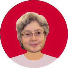
Edition #7: annemie van der Linden, Antwerp
In this episode we discuss mechanisms that the brain employs to clear unwanted waste material whilst we sleep with our former ESMI President and Emerita Professor, Annemie Van Der Linden.
We learn how closely coupled this system, known as the glymphatics, is to blood circulation, and how physical stimulation can enhance glymphatic flow in anaesthetised but not awake mice. Along the way, we learn how curiosity and a wondering mind has led Annemie to use a wide range of models to study the brain, from fish to song birds.
Selected publication:
Glymphatic influx and clearance are accelerated by neurovascular coupling.
Holstein-Rønsbo S, Gan Y, Giannetto MJ, Rasmussen MK, Sigurdsson B, Beinlich FRM, Rose L, Untiet V, Hablitz LM, Kelley DH, Nedergaard M. Nat Neurosci. 2023 Jun;26(6):1042-1053. doi: 10.1038/s41593-023-01327-2.
About the Guest
Annemie Van Der Linden is Professor Emerita and Founder of the Bio-Imaging Lab at the Department of Biomedical Sciences in the University of Antwerp. Annemie received her PhD in Biology in 1989. She became emerita in January 2022. She is the founder and was longstanding director of the Bio-Imaging Lab.
The core interest of her lab and her lifetime research passion has been high resolution MRI of the brain focusing on neurodegeneration, neuroplasticity and ageing using small rodents and songbirds as model systems.
Annemie and her colleagues developed an MRI toolbox to study neurodegeneration, -modulation and -plasticity that has led to many international collaborations and publications. Her team pioneered (f)MRI in tiny songbirds as a remarkable model for vocal learning and neuroplasticity.
She published 300 peer-reviewed publications of which 45 on the use of MRI in songbirds.
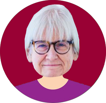
Edition #6: Elisabeth de Vries, Groningen
In this episode we talk about nectin-4 – an emerging biomarker for cancer diagnosis and therapy – with one of the most influential and distinguished scientists in medical oncology, Elisabeth de Vries.
We talk about the first-in-human study of a radioligand that can target nectin-4 and learn what medical oncologists need to get out from imaging data so that these powerful tools continue to make an impact on patient outcomes.
We learn about the importance and best practices for robust clinical trials and the hurdles to both design and finance them.
Along the way, we get a glimpse into Elisabeth’s career path and how her research combines nuclear with optical imaging modalities as methods to improve patient diagnosis and treatment.
Selected publication:
First-in-human study of the radioligand 68Ga-N188 targeting nectin-4 for PET/CT imaging of advanced urothelial carcinoma.
X. Duan, L. Xia, Z. Zhang, Y. Ren, M. G. Pomper, S. P. Rowe, X. Li, N. Li, N. Zhang, H. Zhu 6, Z. Yang, X. Sheng, X. Yang. Clin Cancer Res. 2023 Apr 24; CCR-23-0609. doi: 10.1158/1078-0432.CCR-23-0609.
About the Guest
Elisabeth is a world-renowned Professor of Medical Oncology at the University Medical Centre Groningen. She is involved in patient care, teaching and research. Her research lines aim to increase the sensitivity of tumours to anticancer drugs, and she uses imaging techniques to support this.
She actively promotes a multidisciplinary approach with close interactions between the laboratories and clinic as crucial prerequisite to improve prospects for cancer patients.
She received numerous awards and grants and is a member of the Royal Academy of Arts and Sciences (KNAW), Fellow of the European Academy of Cancer Sciences, and (co)-authored >840 publications (H-index: 137).
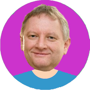
Edition #5: Bernd Pichler, Tübingen
In this episode we learn how a new lensless microscope, costing just $100 (!), can be used to evaluate radiotracer retention in cells, and discuss potential applications for this revolutionary new technology with our famous guest Bernd Pichler. We discuss how this, and other technologies, could be used to assess the heterogeneous cell populations of the tumour microenvironment and identify cell senescence using new radiotracers developed in Bernd’s lab.
Selected publication:
Development of a Lensless Radiomicroscope for Cellular-Resolution Radionuclide Imaging
Justin S. Klein, Tae Jin Kim and Guillem Pratx. J Nucl Med 2023 Mar; 64(3):479-484. doi: 10.2967/jnumed.122.264021. Epub 2022 Sep 15
About the Guest
Bernd is Chair of the Department of Preclinical Imaging and Radiopharmacy and Director of the Werner Siemens Imaging Center at the University of Tübingen, Germany. Moreover, he is the Dean of the Faculty of Medicine of the University of Tübingen.
Bernd works in the field of PET and PET/MR imaging science since more than 15 years and pioneered the development of preclinical and clinical PET/MRI. He performed research at the TU Munich, the MPI for Physics in Munich, UC Davis USA and the University of Tuebingen.
His lab is focussing on interdisciplinary basic research in biomedicine with the use of state-of-the-art imaging technologies. This includes multi-modality imaging in oncology, immunology, and neurology as well as the development of new imaging technologies and innovative imaging probes. In recent years, he has published widely on the preclinical as well as clinical implementation of PET/MR imaging.
Bernd is not only one of the most prominent figures in this wonderful field of research, but a role model, mentor, visionary, and friend to many. He was President of ESMI in 2015 and is certainly still a driving force for the development of this vivid society.
Edition #4: Zaver M. Bhujwalla, Baltimore
In this episode we discuss how targeting fibroblasts rather than tumour cells may be an effective strategy for both surgical guided resection and as an anti-cancer therapy with Tim’s “academic grandmother”, Zaver Bhujwalla. We discovered Zaver’s roots as a physicist and mathematician, learned about her (unpredictable) interest in cancer metabolism and how she combines tools to develop a powerful in vitro diagnostic test.
Selected publication:
Design and characterization of fibroblast activation protein targeted pan-cancer imaging agent for fluorescence-guided surgery of solid tumors
Mukkamala R, Lindeman SD, Kragness KA, Shahriar I, Srinivasarao M, Low PS. J Mater Chem B. 2022 Mar 23;10(12):2038-2046. doi: 10.1039/d1tb02651h.
About the Guest
Zaver Bhujwalla serves as Director of the Division of Cancer Imaging Research, Vice-Chair of Research, and Director of the Molecular Imaging Center and Cancer Functional Imaging Core at the Johns Hopkins University School of Medicine in Baltimore.
Her work is dedicated to the applications of molecular and functional imaging to understand and target cancer and the tumour microenvironment. With over 200 publications in the field, Zaver is certainly one of the pioneers and visionaries who have significantly shaped the field and inspired and mentored generations of (future) imaging scientists. Read more about her work/lab here.
Edition #3: Clemens W.G.M. Löwik, Rotterdam
In this episode we discuss and learn about Bioluminescence imaging and modern tools for multiplexed imaging of different colors, tackling a major challenge in the field. Our guest is one of the pioneers on multicolor BLI. We talk about innovative approaches for unmixing multiple luciferase-luciferin pairs and quantitative analyses of bioluminescent mixtures, enabling serial tracking of heterogeneous cell populations. During the course of our discussion we learn how such methods will help advance BLI to cover new exciting regimes, but also spill over to the wider optical imaging. If you’re interested in learning all about the exciting developments in the field – listen carefully!!

Selected publication:
Multiplexed bioluminescence imaging with a substrate unmixing platform
Caroline K Brennan, Zi Yao, Anastasia A Ionkina, Colin M Rathbun, Buvaneshwari Sathishkumar, Jennifer A Prescher. Cell Chem Biol. 2022 Nov 17;29(11):1649-1660.e4.
doi: 10.1016/j.chembiol.2022.10.004. Epub 2022 Oct 24.
Edition #2: Simon Cherry, UC Davis
In this episode we discuss (and learn a lot about!) positronium imaging with the co-inventor of Total Body PET, Simon Cherry. We talk about the physics behind positronium formation, and how it might provide further biological information than standard positron emission tomography (PET) imaging. We learnt about Simon’s journey that led him to change the field of (clinical) PET imaging and what new innovations he has up his sleeve… and if you are looking for an area of research just waiting to be explored – listen carefully!
Selected publication:
Positronium imaging with the novel multiphoton PET scanner
P Moskal, K Dulski, N Chug, C Curceanu, E Czerwiński, M Dadgar, +30, and W. Wislicki. Sci Adv. 2021 Oct 15;7(42):eabh4394. doi: 10.1126/sciadv.abh4394. Epub 2021 Oct 13.
About the Guest
Simon R. Cherry is a biomedical engineer, and a Distinguished Professor at University of California, Davis. The Cherry lab investigates new technologies and instrumentation techniques in the field of nuclear and optical imaging. Areas of active research include the development of new and improved imaging technologies, the design of novel contrast agents and imaging probes and their application in molecular diagnostics and therapeutics. Simon is the co-inventor of the “EXPLORER” scanner: a high sensitivity, total-body PET system with a 2 meter axial field-of-view.
Edition #1: Jolanda de Vries, Nijmegen
Selected publication:
In vivo imaging of nanoparticle-labeled CAR T cells
by L. Kiru, A. Zlitni, A.M. Tousley +9, and H.E. Daldrup-Link.
About the Guest
Jolanda is head of the Department of Tumor Immunology at the Radboud Institute for Molecular Life Sciences. Her main focus of research is on dendritic cell (DC) biology and on translational immunology. She belongs to the pioneers that translated dendritic cell biology into clinical practice. The first clinical phase I/II studies in which cancer patients were vaccinated with DC loaded with tumor-specific peptides date back to 1997.
Edition #0 & How the Idea Grow: Bertrand Tavitian, Paris
When we recorded an interview with Bertrand on Zoom and watched it afterwards, it became quite obvious that it was… a podcast format! No one wants to sit in front of the screen where there is nothing to see but so much to hear. Thus, enjoy the 6-minutes birth of the ESMI Podcast:

selected publication:
18F-FDG brain PET hypometabolism in patients with long COVID
by E. Guedj et al.
“I go for the unexpected or expected but not done before and then – little by little – you start getting excited (…)”
Download/listen to short interview
Bertrand Tavitian is Professor of Radiology and Medical Imaging. He is the Director of the Imaging Research Laboratory at the University of Paris.
Bertrand is one of the founding fathers of the ESMI and its first President. With his dedication for imaging science, his enthusiasm, and admirable curiosity that goes far beyong “his” field of expertise, Bertrand is an inspiration and mentor for so many scientists in the field.
Guests & Editions
- #18: Metin Sitti, Istanbul
- #17: Rodney Hicks, Melbourne
- #16: Markita Del Carpio Landry, Berkeley
- #15: Vasilis Ntziachristos, Munich
- #14: Peter Scott, Michigan
- #13: Fabian Kiessling, Aachen
- #12: Elisa E. Konofagou, New York
- #11: John Ronald, London ON
- #10: Kevin Brindle, Cambridge
- #9: Mikhail Shapiro, Pasadena
- #8: Jason Lewis, New York
- #7: Annemie Van Der Linden, Antwerp
- #6: Elisabeth de Vries, Groningen
- #5: Bernd Pichler, Tübingen
- #4: Zaver Bhujwalla, Baltimore
- #3: Clemens WGM Lowik, Rotterdam
- #2 Simon Cherry, UC Davis
- #1 Jolanda de Vries, Nijmegen
- #0: Bertrand Tavitian, Paris